Coral Fragments Recover After Tissue Fusion Transfers Beneficial Algae
In lab experiments, bleached coral regained color after fusing with healthy genetic matches, offering a research lead rather than a reef-wide fix.

A bleached coral fragment regained color within nine weeks after it was placed in contact with a healthy, genetically identical fragment. The lab result points to a possible way to support recovery after bleaching: fused coral tissues allowed beneficial algae to move into the depleted fragment. It offers a research direction, though it does not show that the approach can restore reefs at scale.
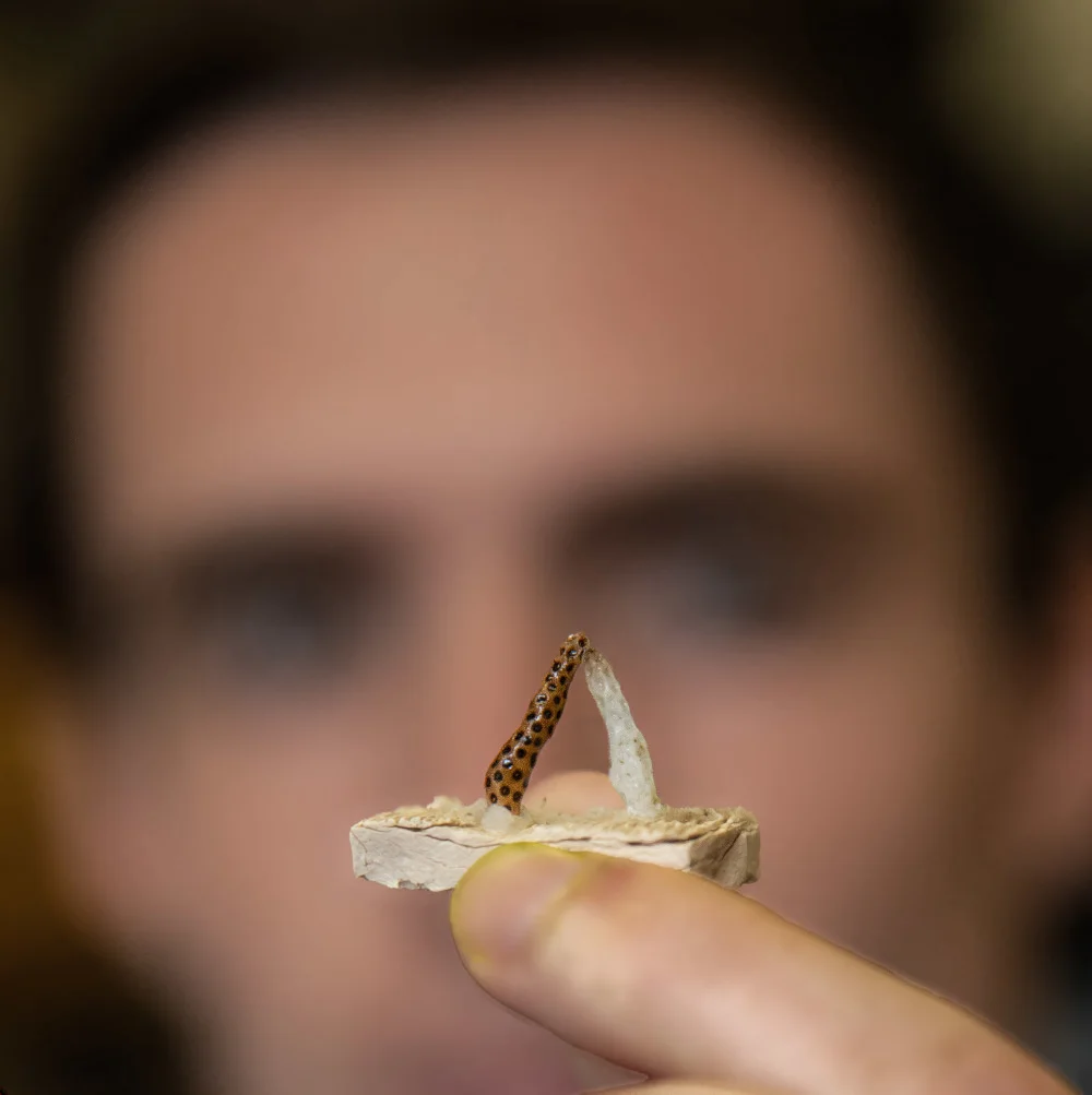
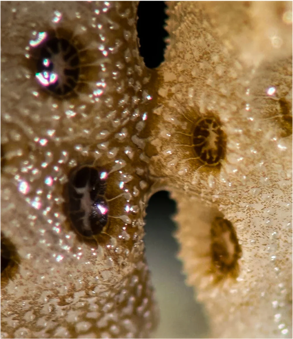
The study, led by Stanford doctoral student Danny Collins, is documented in research published in Science Advances. Its photographs make the change visible: a pale, branching fragment touches a smaller, more colorful one, and later the pale coral has regained color. The point where the two meet becomes both the scientific focus and the visual center of the sequence.
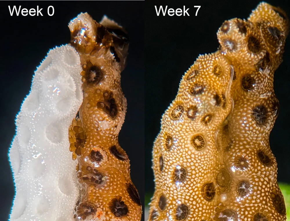
“We thought that some transfer could take place, but we never would have expected it to be so fast, clear, and beautiful,” Collins wrote in an email. The speed and clarity surprised the researchers. The images also give viewers a direct way to follow what happened, without needing to know coral species or experimental methods first.
Collins brought experience in visual and media studies as well as marine science to the work. He said he kept a broad audience in mind as he planned the study and its images. “I wanted everyone to be able to understand our study in just one image and have them say, ‘Wow, I get it.’” The close framing shows the coral’s texture and the contrast between the fragments, then lets the later photograph answer the question raised by the first.
A different kind of before and after
Environmental photographs often show decline: a forest cleared, a glacier diminished, or a reef stripped of color. Those records document damage and help people see what has changed. This sequence moves in the other direction. Its second image shows recovery, making visible not only what bleaching can take away but also a process that may help some coral regain what it lost.
That shift changes the question a viewer might ask. Alongside “What happened here?” comes “What helped?” The answer in this experiment involved contact between genetically identical fragments, tissue fusion and the movement of beneficial algae. The photographs invite interest in the mechanism while retaining the evidence of the original injury.
The visual metaphor is hard to miss. One coral appears to help another, and the contact can call to mind how people support each other through illness or exhaustion. But the fragments’ genetic match and their tissue fusion are biological facts, not evidence of compassion. The sense of an embrace belongs to the viewer. As Collins put it, “I find this represents the power and importance of contact, an idea that can be extended well beyond corals.”
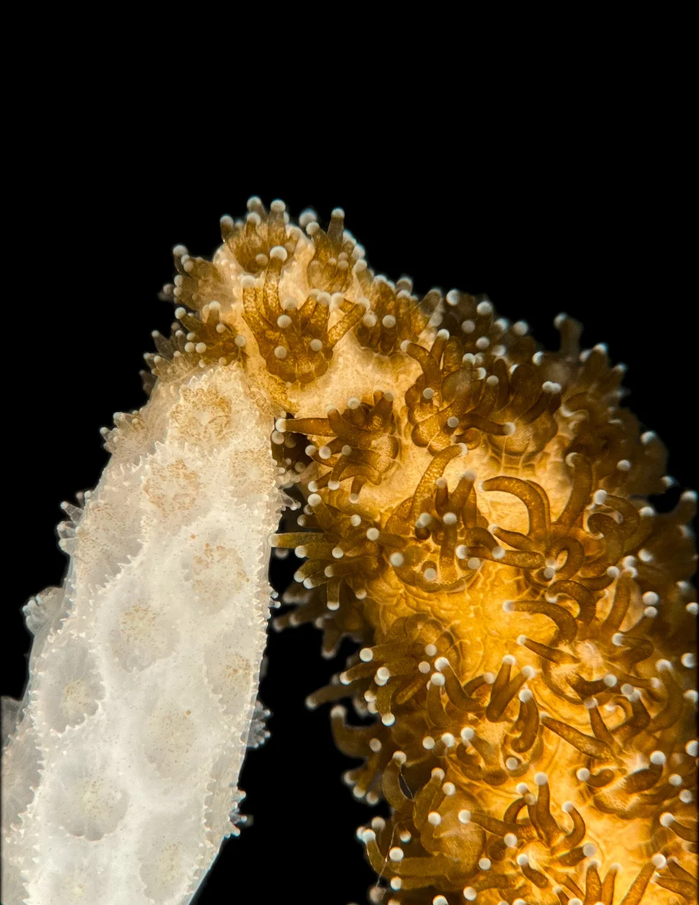
Understanding the mechanism doesn’t erase the emotional force of the image. Tissues connect; algae move; color returns. The beauty and the scientific explanation can sit together, each giving the other more meaning.
What the experiment can—and can’t—show
A compelling pair of photographs can travel farther than the qualifications attached to a research paper. Here, the key limit is that the experiments took place in a lab. Collins cautioned that the work has not solved coral bleaching and that the ocean is still getting warmer. The result opens up possibilities for recovering coral that has already bleached, but it does not establish that the technique will work in reef conditions or at the scale reefs need.
Supplying a healthy, genetically identical partner for every bleached coral would not be feasible at that scale. The experiment identifies a process worth investigating; more work is needed to understand where it may be useful and what conditions recovery requires. For households, coastal communities and businesses that depend on healthy reefs, the practical significance remains uncertain. The study provides a reason to ask what support coral needs after bleaching, not a promise that a single intervention can reverse reef decline.
The pale fragment also challenges a quick judgment about what is beyond help. A coral can be under severe stress while still retaining the capacity to recover under some conditions. That distinction matters in conservation, where assessments made after injury can shape which places or organisms receive attention. The result gives researchers a concrete question to pursue: what can still be done, under what conditions, and with what chance of success?
Recovery begins with an open question
The photographs compress a much longer effort. Researchers maintained the experiment, watched the fragments change, recorded what they saw and examined the evidence with colleagues. Collins described the project as growing from “a simple experiment” into work full of surprises. The finished sequence can make the outcome seem straightforward, but its second image was not guaranteed at the start.
That uncertainty is familiar in restoration. People often begin work after a place has already changed, with incomplete knowledge of what can be brought back. Past conditions can help reveal what has been lost, but they cannot by themselves determine what a damaged place can support in the future. Establishing that requires evidence, local knowledge and choices about priorities. A photograph can make a result visible; it cannot settle those questions.
There is also no image of this coral before it bleached. The sequence starts with damage already present, then asks what happens next. That makes its recovery useful without suggesting that the injury has disappeared or that the original condition can be fully restored. The first panel remains part of the record. The second shows a new outcome that followed an intervention.
For Collins, the discovery began with a question whose answer was unknown. His team found that contact between matched coral fragments could lead to tissue fusion and the transfer of beneficial algae, followed by recovery within weeks in the lab. The result doesn’t tell researchers how the next experiment will turn out. It gives them evidence to shape the next question.
Keeping attention on the work
For people who care about ocean conservation, the photographs can be a starting point for learning about both the finding and its limits. The useful follow-through is to ask what further experiments need, how research can be sustained, and what other conditions support reef recovery. An image can create interest; continued explanation helps turn that interest into understanding.
The paired panels offer a moment of relief, but the original injury remains visible. Their value lies in showing that damage need not be the only possible ending for an individual coral fragment. Recovery was observed after a specific lab intervention. Whether that process can inform efforts in the ocean remains an open question—and a reason to keep studying what happens where the fragments meet.

Previously in The Revelator
Ghost Reefs of 2083: The Paleontology of Color (A Speculative ‘Fiction’)
The post When ‘After’ Means Alive appeared first on The Revelator.